'SEM-to-TEM technique' for higher resolution imaging
One of the drawbacks of SEM blockface imaging is its limitation to magnifications of about 10kx. Due to the similar specimen processing however, SEM blockface imaging can be of tremendous help to facilitate higher resolution TEM studies.
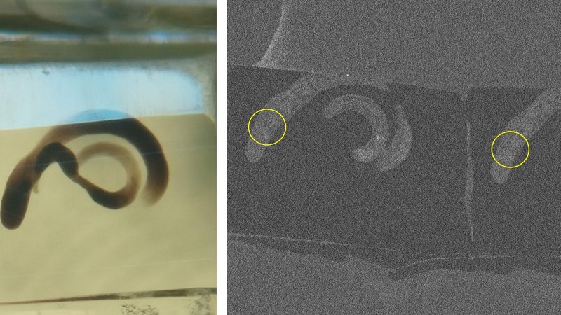
In the ‘SEM-to-TEM technique’, a whole resin block is mounted on a SEM stub. After sectioning into the sample, the block surface is imaged in the SEM. This makes it very easy to get an overview over a larger piece of tissue and to locate a small, specific region of interest. The SEM line scan function can then be used to mark this ROI on the resin, which in turn can guide the trimming of the block in the ultramicrotome. Ultrathin sections of the specific ROI can then be cut and used for TEM imaging at resolutions much higher than those possible by SEM blockface imaging.
In a study conducted by Stefan Koestler (now University of Birmingham) and Rob White (Dept. of PDN, Emeritus) Drosophila testes were labelled in a defined area with a DAB (diaminobenzidine) precipitate using the laser of a confocal microscope (method modified from https://doi.org/10.1126/science.aag002). After subsequent processing of the testes for EM, the 'SEM-to-TEM' technique was used to find and isolate the DAB-labelled area and to prepare sections for TEM tomography studies.
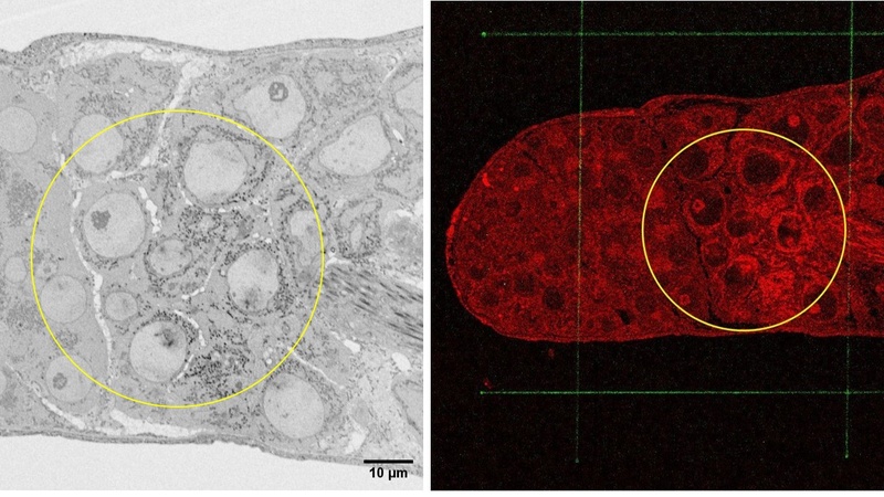
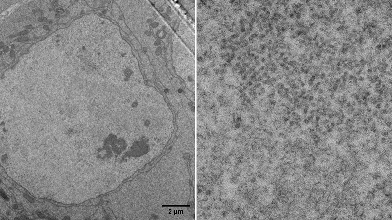
Left: SEM blockface image of the tip of the Drosophila testis at higher magnification. The area of DAB precipitation is recognizable by the darker nuclear and mitochondrial staining. Right: Overlay of the SEM backscatter image (red) with the SE-image of the line scanning marks (green) after marking. The DAB region is denoted by the yellow circle.
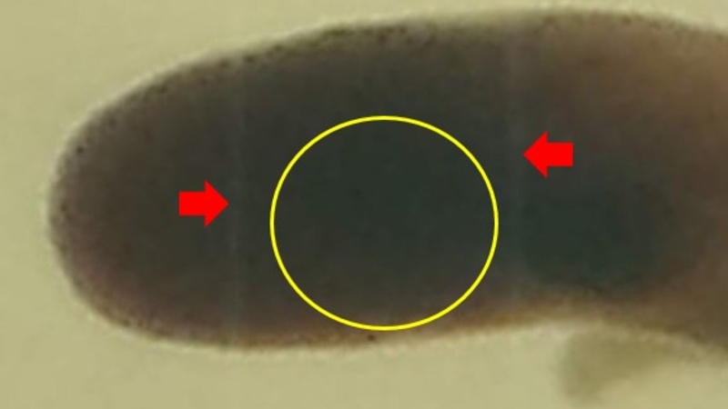
Image of the marked resin block in the ultramicrotome. The line scan marks on the resin are denoted by the red arrows and can be used as guidance to trim the block to a specific area of interest (yellow circle).