
Specifications
Specifications of multiview light sheet microscope
The multiview light sheet microscope is equipped with four excitation laser lines and a set of filters to match some of the most common fluorescent fluorophores.
Fluorescent proteins like GFP, YFP, RFP, mCherry as well as dyes with similar spectra can be imaged.

Principle layout of the multiview light sheet microscope. Excitation light enters from the upper right corner and the sample is mounted in the sample chamber visible in the lower part of the picture.
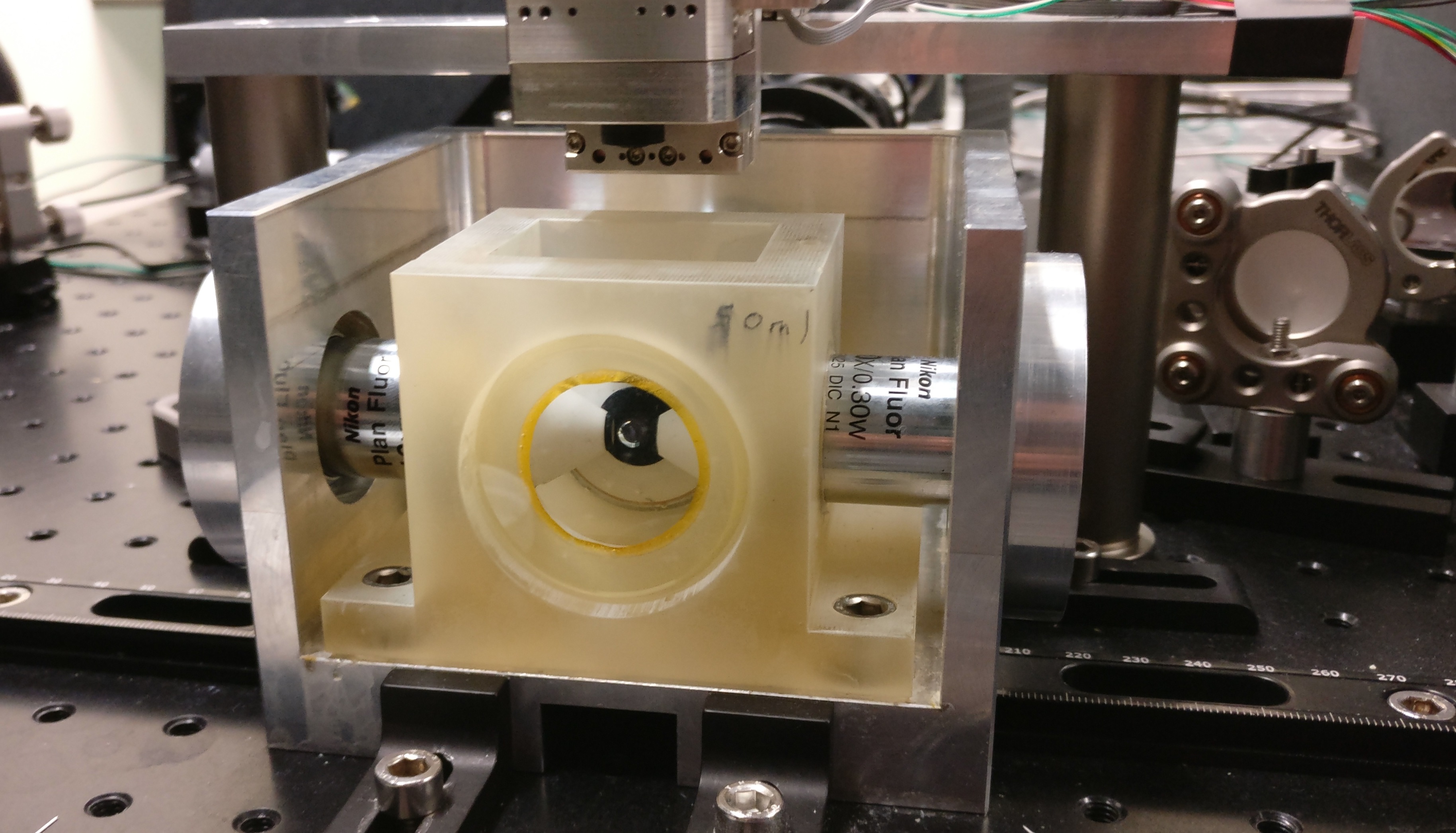
A close up of the sample chamber of the microscope. The sample is mounted in a capillary in the movable sample holder seen above the chamber. The samples are immersed in water based solutions and imaged with water dipping objectives.
Excitation wavelengths:
- 445 nm
- 515 nm
- 488 nm
- 561 nm
Emission filters
- 460-500 nm (480/40)
- 500-550 nm (525/50)
- 525-565 nm (545/40)
- 570-640 nm (605/70)
- 620-695 nm (670/50)
Objectives:
- Nikon Plan Fluo 10x (excitation)
- Nikon APO LWD 25x (emission)
Detection: Hamamatsu Orca Flash 4 camera
Sample movement provided by piezomotor driven stages (http://www.nanos-instruments.de)
The microscope is controlled by a custom designed software. The software as well as the user interface are implemented using Labview in order to allow for maximum flexible and easy user operation.