
Analysis of self-organising aggregates of zebrafish retinal cells
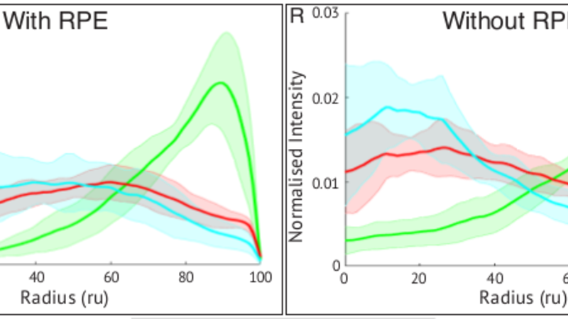
For the study of formation of retinal layers, the fluorescence patterns in images of cultured dissociated Zebrafish retinal progenitors were extracted and compared.
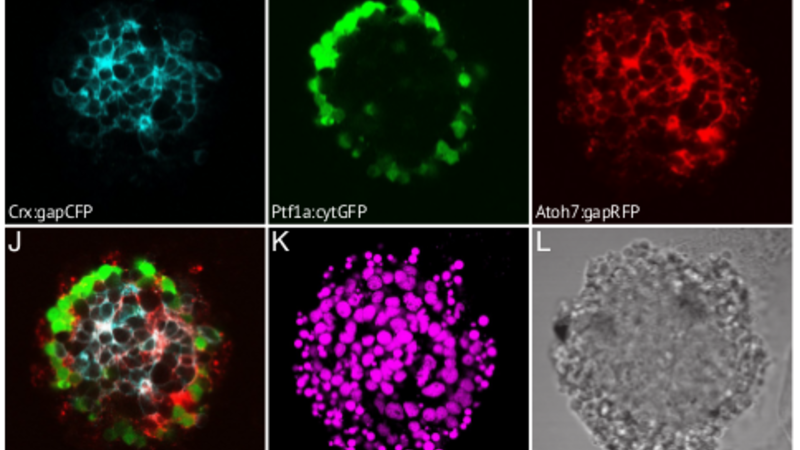
Quantitative measures of lamination were computed from the fluorescence images , which were then used to show that Müller glia, but not RPE cells, are essential for this process.
Collaboration with Megan Eldred (Harris group)
Reference:
Eldred MK, Charlton-Perkins M, Muresan L, Harris WA. - Self-organising aggregates of zebrafish retinal cells for investigating mechanisms of neural lamination, Development, 144(6):1097-1106, 2017. doi: 10.1242/dev.142760