Space-varying deblurring of lightsheet microscopy images
Deconvolution, computational microscopy
This project is a collaboration between Dr Bogdan Toader, Dr Leila Muresan (CAIC), Prof Carola Schoenlieb, Dr Yury Korolev (DAMTP) and Dr Jerome Boulanger (MRC-LMB).
Image formation in light microscopy images leads to space variant blur and intensity roll off due to the due to the interaction between the illumination and detection point spread functions (PSF). In this project, we have developed a flexible and accurate model of the imaging process for lightsheet microscopy. This forward model goes beyond the limitation of spatially constant PSF assumed by traditional deconvolution algorithms. We also have incorporated a rigorous noise modelling that enable automatic parameter tuning. The optimization problem is then solved using efficient algorithms that can be seamlessly ran on GPU.

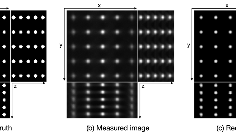
In Figure 1, we applied our method to a simulated 3D stack of beads, where we see clearly the effect of the illumination on the PSF — the beads on the left and right hand sides of the image are more blurry and elongated in the z direction than the ones in the centre. This effect is successfully removed in the reconstructed image.
