
Single color STED spermatocytes
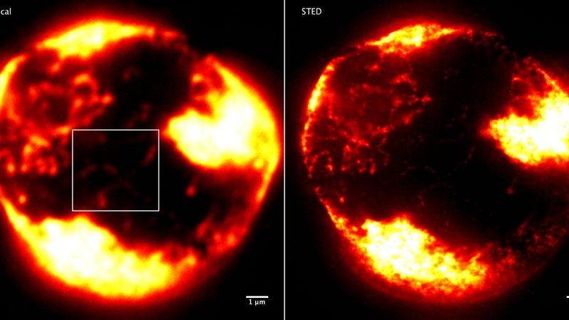
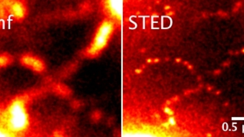
Drosophila primary spermatocyte nucleus labeled with anti-histone (STAR Red) showing the organization of chromatin blocks (chromosomes) and, at higher power, the structure of individual chromatin fibres.
Comparison of standard confocal imaging (left panels) and STED imaging (right panels)
Images taken in collaboration with Rob White, Department of Physiology, Development and Neuroscience.