
Backscatter imaging
Backscatter imaging
Backscatter (BSc) imaging is a second imaging modality available in SEM. While SE-imaging reveals the surface topography of our sample, BSc-imaging can provide information as to its composition. Elements with a high atomic number yield more backscatter electrons than elements with a low atomic number and hence appear bright in the image. Imaging a sample in BSc-mode provides therefore a Z-contrast image indicating differences in elemental composition. Samples for BSc-imaging are imaged uncoated or coated with a layer of carbon to improve sample conductivity.
The Verios 460 is equipped with a range of backscatter detectors: the ETD and TLD detectors can be run in both SE-mode or BSc-mode. The CBS (concentric backscatter) detector is a dedicated BSc detector and its concentric segmentation allows to select for electrons scattered by the sample at different angles. The mirror detector (MD) and the In-column detector (ICD) are also dedicated BSc detectors, sitting increasingly high inside the SEM column, and can be used to obtain increasingly ‘pure’ Z-contrast images of material samples. BSc-imaging is frequently used in conjunction with EDX analysis.

Carbon nanotubes imaged in SE-mode (left) showing their smooth surface topography. The image on the right is an overlay of this SE-image with the BSc image (red) revealing areas of high Z inside the CNTs. EDX showed this to be the iron catalyst used during CNT synthesis (Prof. Slawomir Boncel, Silesian University of Technology, Poland; DOI: 10.1021/acsbiomaterials.6b00197).
However, backscatter imaging can also be used to image metal sputter coated samples to reveal structures at greater depth in a sample.
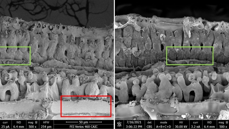
Partial fracture of freeze-dried wheat leaf. On the left, imaged in SE-mode using the ETD detector at 2 keV and 25 pA detailing mostly the surface topography of the sample. On the right, the leaf was imaged in BSc-mode using the CBS detector at 30 keV and 3.2 nA reaching more structures in the depth of the sample, e.g. intracellular chloroplasts (green square) or penetrating the surface wax layer (red square) to reveal the underlying epidermis cells (Karin Mueller, CAIC).