
Cryo-SEM
Cryo-SEM at CAIC
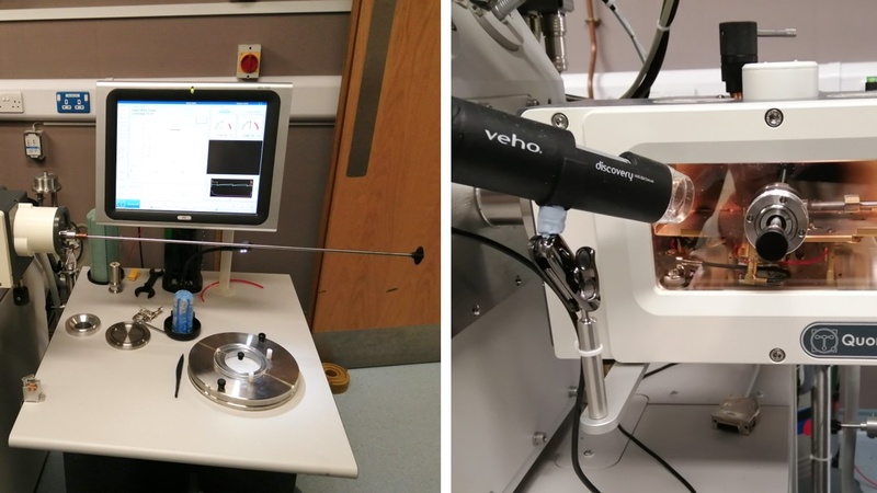
Our Verios 460 SEM is equipped with a Quorum PP3010T cryo-SEM preparation system. Cryo-SEM allows imaging of soft, hydrated biological or materials samples in a frozen state. One of the major advantages of cryo-SEM is that samples can be imaged with minimal preparation without the need of any prior fixation or dehydration steps; a disadvantage is that sample throughput is fairly low compared to other methods. Specimens are mounted on an appropriate cryo-SEM shuttle, are plunge-frozen in slushed liquid nitrogen and then transferred to a cooled prep stage. Once on the prep stage, samples can be fractured, sublimed to remove any residual ice, and coated to improve contrast and conductivity (CAIC routinely uses a platinum target). Then, the sample is transferred onto the SEM cryo-stage for imaging. As cryo-SEM requires about 2h of run-up and run-down time of the system apart from the actual imaging time, we recommend half-day or whole-day bookings.
Some examples of cryo SEM:

Geranium petal, surface plus fracture at the petal base (Karin Müller, CAIC, Cambridge).

Pitcher plant, leaf surface with scales (Karin Müller, CAIC, Cambridge).

Horsetail, stomata and wax on the leaf surface (Karin Müller, CAIC, Cambridge).

Plantain stem (fractured) showing cell walls and cell interior with various organelles and delineating membranes (Karin Müller, CAIC, Cambridge).

Curdled milk (fractured) showing round milk fat globules within a water/solute matrix (Karin Müller, CAIC, Cambridge).

Blue cheese (fractured) showing Penicillium spores (Karin Müller, CAIC, Cambridge).