
Laser ablation
Laser ablation - SEM blockface imaging
Embryos must undergo a dramatic change in shape early on in development in order to transform from a collection of cells into a fully functional organism. A crucial step in this process is the elongation of the head-to-tail embryonic axis. A study using a combination of light-mediated cell ablation and quantitative analysis of tissue and embryo morphogenesis found that coordination between anterior notochord cell expansion and posterior addition to the notochord from a pool of progenitors generates a force that facilitates the elongation of the embryo head-to-tail axis (Susannah McLaren, Group of Ben Steventon, Department of Genetics, University of Cambridge; https://doi.org/10.1242/dev.199459).
To enhance the understanding of the tissue and cellular damage caused by the ablation, a somewhat ‘atypical’ CLEM experiment was performed where the ablated regions were subsequently imaged in greater detail by SEM blockface imaging. This study showed that laser ablation leads to cell fragmentation in a very accurate fashion with a sharp delineation between unaffected and damaged tissues.
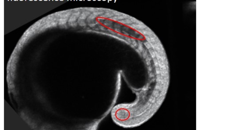
Confocal image of a zebrafish embryo, the two ablations, a large area in the notochord and a small area in the tail bud, are highlighted in red.
Following the ablation in a confocal laser microscope, the zebrafish embryos were processed for electron microscopy, sectioned and viewed by SEM blockface imaging with the aim to find the ablated areas.
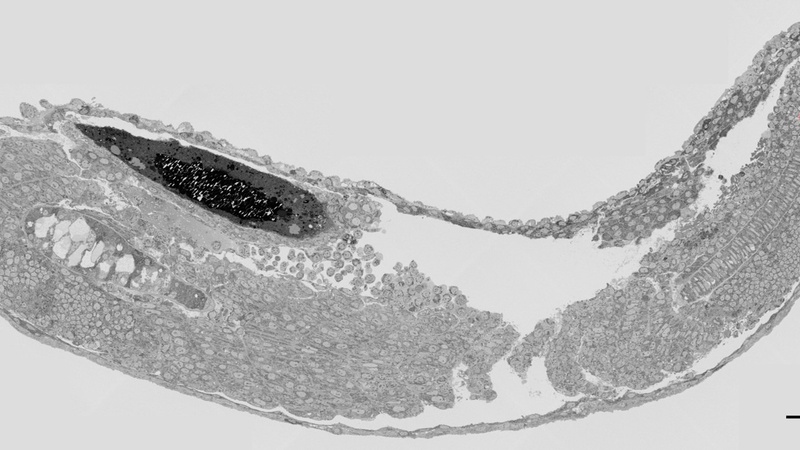
Low resolution map of a zebrafish embryo, the ablated area in the tail bud is circled in red. The larger ablation in the notochord, apparent in the confocal image, is not visible in this section as it lies more anteriorly along the axis at a different section depth.
Once an ablated area is identified, the morphology of the tissue can be mapped in the SEM at increased magnifications.
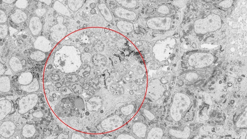
Medium resolution map of the zebrafish tail bud. The tissue area damaged by the laser is circled in red.
The cell damage caused by the ablation becomes apparent at higher magnification. The close vicinity of normal versus damaged tissue is a testament to the accuracy of the laser ablation technique.
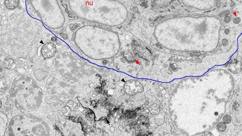
The blue line denotes the border between the ablated/damaged tissue area (bottom) and the normal, undamaged tissue (top). The red markings show undamaged, oval nuclei (nu) with a ‘tight’ nuclear membrane, elongated mitochondria with a dark matrix (arrowheads) and Golgi membrane stacks (arrow). In contrast, the black markings in the ablated area show round nuclei with condensed chromatin and dilated nuclear membranes and the mitochondria have become enlarged and rounded with dilated cristae and a pale matrix (arrowheads). The normal cellular structure is highly disrupted and dark-stained areas of collapsed cell debris (arrow) are apparent.